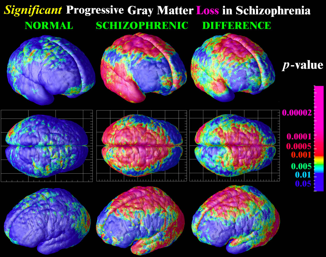
the idea is this |
|
Results 1 to 8 of 8
 5Likes
Thread: schizophrenia and lucid dreams
Hybrid View
-
08-03-2011 05:54 PM #1Member


- Join Date
- Jul 2011
- LD Count
- 2 x pie^squared
- Gender

- Location
- on the edge of reality
- Posts
- 194
- Likes
- 31
- DJ Entries
- 5
 schizophrenia and lucid dreams
Last edited by hpnfreak; 08-03-2011 at 05:57 PM.
dream goals:
[ ] - sail with the Black Perl to Hoghwarths and show my magic wand to Hermione aaarrrrrrrrr
[ ] - turn into Chuck Norris,kick Justin Bieber into a large pit, while shouting "shut the f*ck uuuuuuuuuupppp"
[ ] - meet the Sasquatch and ask him to pick my stocks, then crash the economy
[ ] - find my virginity to avoid the piranhas on the escalator and get probed by aliens
-
08-05-2011 08:22 AM #2Member


- Join Date
- Jul 2011
- LD Count
- 2 x pie^squared
- Gender

- Location
- on the edge of reality
- Posts
- 194
- Likes
- 31
- DJ Entries
- 5
so this thread wasn`t a good idea at all
dream goals:
[ ] - sail with the Black Perl to Hoghwarths and show my magic wand to Hermione aaarrrrrrrrr
[ ] - turn into Chuck Norris,kick Justin Bieber into a large pit, while shouting "shut the f*ck uuuuuuuuuupppp"
[ ] - meet the Sasquatch and ask him to pick my stocks, then crash the economy
[ ] - find my virginity to avoid the piranhas on the escalator and get probed by aliens
-
07-22-2012 09:58 AM #3Member Achievements:


- Join Date
- Mar 2008
- LD Count
- In DV +216
- Gender

- Location
- In a Universe
- Posts
- 994
- Likes
- 1139
- DJ Entries
- 88
.../Lucid dreaming – when you are aware you are dreaming – is a hybrid state between sleeping and being awake. It creates distinct patterns of electrical activity in the brain that have similarities to the patterns made by psychotic conditions such as schizophrenia/...
Source: New Links Between Lucid Dreaming And Psychosis Could Revive Dream Therapy In Psychiatry
-
07-21-2012 05:39 PM #4Member Achievements:


- Join Date
- Jul 2012
- LD Count
- 112
- Gender

- Location
- Pittsburgh PA
- Posts
- 230
- Likes
- 56
- DJ Entries
- 13
I met a schitzophrenic on the internet who said when he was 7, he could capture and fight real life looking pokemon. He said that the extremely severe schitzophrenics don't even understand the concept of dreaming but the less severe ones can always tell when they are dreaming because they learn how to difrentiate the things they see between whats actual reality. I asked him if lucid dreaming came easy to him, he see he always knows when he's dreaming unless it's a nightmare.
-
07-23-2012 10:00 PM #5
That's actually pretty interesting, the ability to distinguish dreams and dream-like hallucinations just arose out of a necessity to be able to function in normal reality.... I always kind of thought dreaming really just works that way, too. I've never used a single technique to practice getting lucid other than the occasional very weak mantra ("I'll realize that I'm dreaming tonight.") and RCs twice, by accident. It just gets easier every time I get lucid... it becomes simpler to tell the difference between reality and the dream. I think that's really all it takes, you just have to pay attention.
-
07-23-2012 10:16 PM #6Member


- Join Date
- Jul 2012
- Gender

- Location
- UK
- Posts
- 21
- Likes
- 2
I just had to reply to this thread as a medical doctor that has recently studied psychiatry.
Schizophrenia is nowhere near as simple as you suggest - no one knows what causes it. Some have visible brain damage, some have completely normal looking brains. The theory you present about left and right brains may be true for a small subset of schizophrenics, but this is NOT the main theory and no-one has any real idea what's going on.
Schizophrenia is also not one condition. There are many different types of schizophrenia each of which have completely different behaviours.
Some schizophrenics just stay quiet and seem to have no emotions and no drive to do anything. This is not the standard picture of the schizophrenic we see on TV, but it is quite common and very sad because it is almost untreatable. Some people start off like this, some start off with another form of schizophrenia which is treated, but then they eventually become like this.
Some schizophrenics, as you say, are quite obviously crazy and do/say crazy things. But even then, there are lots of different types and they all behave very differently.
If you want real up-to-date information on Schizophrenia, look here:
emedicine.medscape.com/article/288259-overview#showall
This is one resource that doctors (especially in America) use and which is kept up to date and is reasonably detailed.
-
07-23-2012 10:23 PM #7
The original poster isn't going to respond; it seems he was only active for about a month after registering and hasn't signed on for nearly a year since.
Though I must say, if you what you say is accurate then that's quite annoying. Nearly every neurochemical research article ever made that studies schizophrenia doesn't differentiate between different subsets. In other words, combined with what you've said, it's all completely useless. (Not that I thought any differently as it was, since it's all fairly contradictory and inconclusive anyway.)
-
07-23-2012 10:34 PM #8Member


- Join Date
- Jul 2012
- Gender

- Location
- UK
- Posts
- 21
- Likes
- 2
Yep I see. Oh well, at least some people might benefit from learning it's not so simple.
As for the research articles you seem to have read, well, the link I provided has a number of possible theories as to what might be causing Schizophrenia. I'll copy paste the section on Pathophysiology below:
 Originally Posted by eMedicine
Similar Threads
-
Schizophrenia + lucid dreaming?
By kalbaecnailla in forum General Lucid DiscussionReplies: 5Last Post: 08-30-2010, 04:47 AM -
Can lucid dreaming trigger schizophrenia?
By imj in forum General Lucid DiscussionReplies: 35Last Post: 09-08-2009, 05:19 PM -
Lucid Schizophrenia?
By jamous in forum Dream ControlReplies: 16Last Post: 04-15-2008, 05:40 AM -
schizophrenia
By mongreloctopus in forum The LoungeReplies: 27Last Post: 04-07-2006, 07:22 PM -
schizophrenia?
By jrh7r9 in forum General Lucid DiscussionReplies: 6Last Post: 01-18-2005, 04:40 AM


 LinkBack URL
 About LinkBacks


Bookmarks